
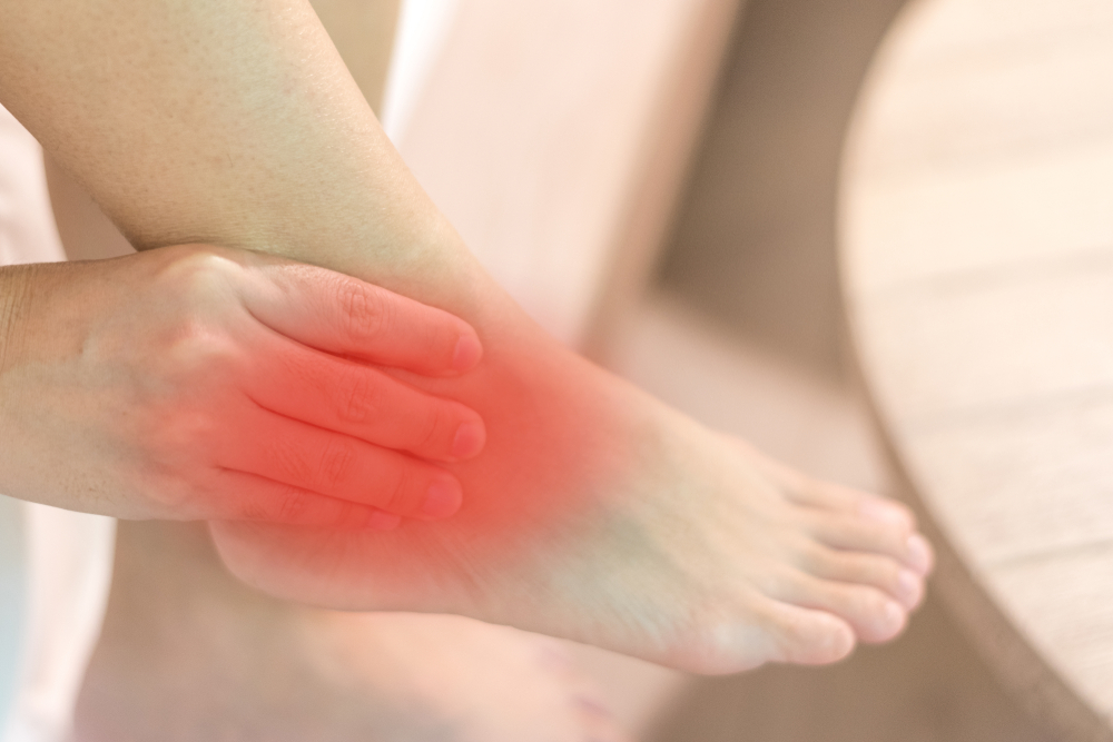
Chronic Foot or Ankle Pain That Gets Worse With Activity and Better With Rest — This Is What Arthritis Feels Like
Osteoarthritis is the most common form of arthritis and one of the most common causes of chronic foot and ankle pain in adults. It develops when the cartilage that cushions and protects the joint surfaces breaks down over time, leaving bone to rub against bone with progressively less protection. The result is pain with movement, stiffness after rest, swelling, and eventually limited range of motion and difficulty with activities that were once effortless.
In the foot and ankle, osteoarthritis most commonly affects three locations: the big toe joint (first metatarsophalangeal joint), the midfoot (the joints of the Lisfranc complex and naviculocuneiform region), and the ankle (tibiotalar joint). Each location produces a characteristic pattern of symptoms and requires a specific management approach. Our podiatrists evaluate and treat foot and ankle osteoarthritis at our Coon Rapids and Golden Valley offices, serving patients across the north and west metro including Maple Grove, Ham Lake, Andover, Champlin, Blaine, Plymouth, Wayzata, Minnetonka, St. Louis Park, and Edina.
Foot and ankle osteoarthritis is manageable at every stage. Cartilage cannot be regenerated, but pain and function can be significantly improved with the right combination of footwear, orthotics, injection therapy, and — when appropriate — surgery. Many patients who believe they are “just getting older” are living with undertreated arthritis that responds well to targeted care.
Call 763-421-7300 | Book online
The Three Most Common Locations
Big Toe Joint (First Metatarsophalangeal Joint)
Arthritis at the first MTP joint is called hallux rigidus or hallux limitus depending on the degree of motion loss. It is the most common arthritic condition of the foot. Pain occurs primarily with push-off and activities that require the big toe to bend upward (stairs, inclines, running). A dorsal bone spur often develops on the top of the joint and is palpable as a bump.
See our Big Toe Arthritis (Hallux Rigidus) page for the complete clinical picture, Coughlin-Shurnas grading, and full treatment discussion for this specific joint.
Midfoot (Lisfranc Complex and Naviculocuneiform Joints)
Midfoot osteoarthritis affects the tarsometatarsal (Lisfranc) joint complex and the naviculocuneiform joints — the region where the long metatarsal bones meet the cuneiform and cuboid bones in the mid-arch area. It is less commonly recognized than ankle or toe arthritis but is a significant source of chronic midfoot pain and functional limitation.
Primary causes: prior Lisfranc injury (sprain or fracture-dislocation) is the most common cause, often developing years after an injury that seemed minor at the time. Severe flatfoot or high-arched foot create abnormal midfoot stress distribution that also accelerates degenerative change.
Characteristic symptoms: diffuse aching across the mid-arch worsened by prolonged standing and walking, relief with rest, pain on direct palpation across the Lisfranc joint line, difficulty walking barefoot on hard floors, and preference for supportive shoes at all times. A dorsal bony prominence may develop.
Management: custom orthotics with stiffened forefoot, carbon fiber foot plates, and targeted steroid injections are highly effective conservative options. Surgical fusion of the affected joints is the definitive treatment for advanced cases.
Ankle (Tibiotalar Joint)
Ankle osteoarthritis is most commonly post-traumatic — meaning it develops as a direct consequence of prior injury to the ankle, most often an ankle fracture or severe ligamentous instability from repeated sprains. This distinguishes it from hip and knee OA, which are primarily age-related wear-and-tear conditions. The average patient with ankle OA is in their 50s and often has a clear history of ankle injury in their 30s or 40s.
Why post-traumatic OA develops: a fracture disrupting the joint surface, or chronic instability from repeated sprains causing abnormal wear patterns, damages joint cartilage permanently. Over the following decade or two, the damaged cartilage progressively deteriorates, the joint space narrows, and bone spurs form around the joint margins.
Characteristic symptoms: deep aching ankle pain during and after activity; morning stiffness lasting more than 15–30 minutes; swelling at the end of the day; reduced ankle motion; difficulty on uneven surfaces; and progressive loss of activities the patient was previously able to do.
Post-traumatic ankle OA is not “just aging.” A patient in their 50s with significant ankle OA after a fracture in their 30s is experiencing the long-term consequence of a specific injury — not age-related degeneration. An accurate diagnosis with weight-bearing X-rays and thorough history is the starting point.
Causes & Risk Factors
Prior joint injury: the most important risk factor for ankle and midfoot OA. Fractures, Lisfranc injuries, and chronic ankle instability from repeated sprains all accelerate cartilage degradation in the affected joint.
Age and cumulative wear: cartilage thins and loses its shock-absorbing properties with age and repetitive loading, particularly in weight-bearing joints. OA prevalence increases sharply after age 50.
Foot structure: flatfoot increases midfoot and Lisfranc joint stress; high-arched (cavus) foot concentrates impact loading and creates rigid jamming of joints. Both accelerate OA in their respective mechanical stress patterns.
Obesity: excess body weight significantly increases the load on all foot and ankle joints with every step. Each pound of body weight multiplies to 3–7 times that force across the joints during walking.
Inflammatory arthritis: rheumatoid arthritis, psoriatic arthritis, and ankylosing spondylitis can cause joint destruction that resembles or coexists with OA. These require systemic treatment in addition to local foot and ankle management.
Osteonecrosis and prior infection: avascular necrosis of the talus or navicular, or prior septic arthritis of the ankle, can cause rapid cartilage loss and secondary OA.
Diagnosis
Weight-bearing X-rays are the foundation of OA diagnosis and staging. The key findings on X-ray are joint space narrowing (reflecting cartilage loss), subchondral sclerosis (bone hardening beneath the cartilage), osteophyte (bone spur) formation at the joint margins, and in advanced cases, subchondral cysts. We always obtain X-rays weight-bearing when possible — non-weight-bearing X-rays underestimate the degree of joint space narrowing.
Weight-bearing X-ray — the primary diagnostic study. Grades severity and guides treatment planning.
CT scan — provides detailed three-dimensional assessment of bony anatomy for pre-surgical planning, particularly for ankle and midfoot fusion procedures.
MRI — used when cartilage integrity or osteonecrosis needs to be characterized beyond what X-ray shows, particularly in early disease or when the degree of cartilage loss will determine surgical candidacy.
Diagnostic injection — a targeted corticosteroid injection into the suspect joint, confirming the source of pain while providing temporary treatment. Particularly useful in the midfoot where multiple adjacent joints can produce overlapping pain patterns.
Treatment at Family Foot & Ankle Clinic
Treatment is matched to the severity of OA, the specific joint involved, and the patient’s functional goals. For most patients, a significant improvement in symptoms and function is achievable without surgery.
Footwear Modification
Appropriate footwear is the most immediately impactful conservative intervention. For foot and ankle OA:
Rocker-sole shoes: the rocker curved sole redirects forefoot roll-through without requiring joint motion, dramatically reducing pain in midfoot and big toe OA. Many athletic shoe brands include a rocker geometry — look for a curved forefoot rather than a flat sole.
High-top or ankle-support footwear: for ankle OA, a shoe or boot with added ankle support reduces tibiotalar joint motion and associated pain during walking.
Stiff-soled shoes: a shoe that does not flex at the forefoot reduces pain in both midfoot and big toe OA by eliminating the bending motion that loads the arthritic joints.
See our Shoe Recommendations Guide for current specific picks.
Orthotics and Joint Support
Over-the-counter orthotics: A semi-rigid arch support can help improve foot alignment and reduce stress across arthritic joints. For arthritis affecting the midfoot or big toe, a full-length rigid carbon fiber insert can limit painful joint motion during walking and often provide substantial relief. Download our Osteoarthritis Patient Guide below for our current recommendations on over-the-counter orthotics and carbon fiber inserts.
Custom orthotics. For patients with significant biomechanical abnormalities, such as flat feet or high arches, or for those who have not achieved adequate relief with supportive footwear and over-the-counter inserts, a custom orthotic provides more precise control of foot mechanics. Joint-specific modifications, including cutouts, extensions, and posting, can further offload arthritic joints and reduce pain. Learn more on our Custom Orthotics page.
Ankle-foot orthosis (AFO) or ankle brace. For hindfoot and ankle arthritis, a custom or semi-custom ankle-foot orthosis (AFO) can reduce painful hindfoot and ankle joint motion while providing stability during walking. Ground reaction AFOs and Arizona braces are well-established options for patients with advanced ankle arthritis who are not yet candidates for surgery or who prefer to delay surgical treatment.
Injection Therapy
Corticosteroid injection: targeted injection into the affected joint provides significant short-to-medium term pain relief and reduces intra-articular inflammation. Particularly effective during acute flares. Used selectively — repeated injections at short intervals can accelerate cartilage loss and are avoided.
Physical Therapy and Exercise
Strengthening the muscles surrounding an arthritic joint reduces joint loading and improves stability. Range of motion exercises maintain what mobility remains. Aquatic therapy is well-tolerated because water buoyancy offloads the joint while allowing movement through its available range.
Surgical Treatment
Surgery is considered when conservative management has been optimized for an adequate period (typically 3–6 months) without sufficient relief, or when the OA is so advanced that conservative care cannot provide meaningful improvement.
Joint fusion (arthrodesis): the gold standard for end-stage ankle and midfoot OA. The joint surfaces are removed and the joint is stabilized with plates and screws until the bones fuse permanently. Eliminates joint pain definitively. The patient loses motion at the fused joint permanently, but the functional result — a stable, pain-free platform for walking — is highly successful. Ankle fusion and midfoot fusion each have high patient satisfaction rates in appropriately selected patients.
Total ankle replacement (arthroplasty): an alternative to ankle fusion in selected patients, particularly those with lower activity demands. Preserves some ankle motion and reduces stress on adjacent joints. Outcomes continue to improve with newer implant generations.
Debridement and bone spur removal: for early OA with significant osteophyte impingement but preserved joint space, arthroscopic or open spur removal restores range of motion and reduces pain without sacrificing the joint.
See our Foot & Ankle Surgery Overview for general information on surgical care at our clinic.
Frequently Asked Questions
Is arthritis in my foot and ankle permanent?
The cartilage loss that has already occurred is permanent — cartilage does not regenerate to a meaningful degree in adults. But arthritis is not uniformly progressive, and many patients maintain stable function for years with appropriate management. The goals of treatment are reducing pain, maintaining mobility and strength, slowing progression where possible, and preserving function for as long as possible before surgical intervention becomes necessary.
My X-ray shows bone spurs and joint space narrowing. Does that mean I need surgery?
No. The degree of arthritis on X-ray does not necessarily directly correlate with symptoms. Some patients with significant X-ray changes have minimal pain and function well with conservative management; others with moderate X-ray changes are substantially limited. Treatment decisions are based on symptoms and functional limitation, not X-ray findings alone. If your X-ray looks severe but you are managing well with conservative care, surgery is not indicated.
I had an ankle fracture years ago. Is my current ankle pain arthritis?
Possibly. Post-traumatic ankle OA typically develops 10–20 years after a significant ankle injury. If your ankle was fractured or severely sprained and you are now experiencing chronic deep ankle pain, stiffness, and swelling, post-traumatic OA is the most likely diagnosis. A weight-bearing X-ray will characterize the degree of joint space narrowing. The earlier post-traumatic OA is identified and managed, the more conservative options remain available.
What is the difference between gout and osteoarthritis in the big toe?
Gout causes sudden, intensely painful, red, hot swelling at the big toe joint from uric acid crystal deposition. Osteoarthritis causes chronic, gradual stiffness and aching with activity, with a progressive reduction in how far the toe bends. Both can affect the same joint, and prior gout flares accelerate OA progression by damaging the cartilage. If you have sudden, severe big toe pain with redness and heat, seek evaluation for gout. If your big toe is chronically stiff and painful with activity, see our Big Toe Arthritis (Hallux Rigidus) page.
Chronic Foot or Ankle Pain That’s Limiting What You Can Do? Let’s Find Out What We’re Dealing With.
Most foot and ankle osteoarthritis responds better to treatment than patients expect. The right combination of footwear, orthotics, and injection therapy can significantly reduce pain and delay or avoid surgery. Weight-bearing X-rays and a clinical examination are the starting point. Book an appointment at our Coon Rapids or Golden Valley office.
Call 763-421-7300 | Book online
Serving Coon Rapids, Golden Valley, Maple Grove, Ham Lake, Andover, Champlin, Blaine, Plymouth, Wayzata, Minnetonka, St. Louis Park, Edina, and all Twin Cities communities.