
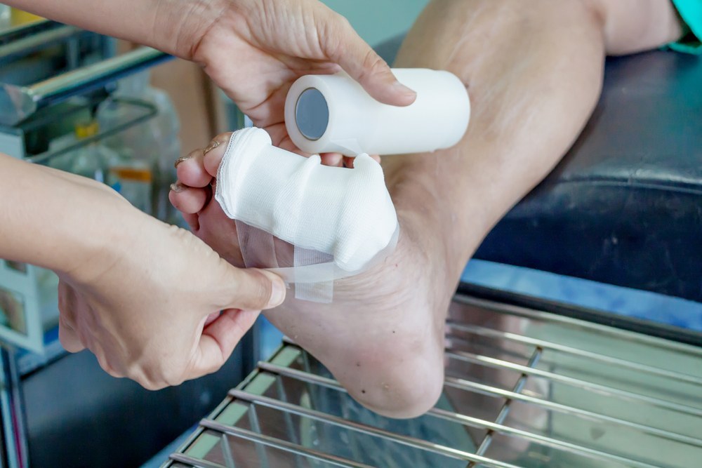
A Foot Wound That Isn’t Healing Is a Medical Emergency in the Making
A cut, blister, or sore on the foot is a minor nuisance for most people. For someone with diabetes, it can be the beginning of a cascade that ends in amputation. Diabetes impairs the two mechanisms the body relies on to prevent foot wounds from becoming life-threatening: sensation, which would normally prompt you to remove the source of injury, and circulation, which is what heals the wound once it exists. Without both, even a small break in the skin can become a deep ulcer, then a bone infection, then a surgical emergency.
Approximately 19%–34% of people with diabetes will develop a foot ulcer during their lifetime. Of those who develop an ulcer, about one in five will ultimately require an amputation of part of the foot or leg. While not every amputation can be prevented, many can be avoided through early recognition, prompt treatment, and ongoing multidisciplinary foot care. Our podiatrists provide diabetic foot wound care and ulcer management at our Coon Rapids and Golden Valley offices, serving patients across the north and west metro including Maple Grove, Ham Lake, Andover, Champlin, Blaine, Plymouth, Wayzata, Minnetonka, St. Louis Park, and Edina.
If you have a foot wound that has not shown meaningful improvement within two weeks, or any wound with redness, warmth, drainage, or odor: call our office today. Same-day and next-day appointments are available for diabetic wound evaluation. Do not wait for a scheduled appointment if the wound is changing rapidly, if red streaking is present, or if you have fever, chills, or are feeling systemically unwell. These are signs of advancing infection and require emergency evaluation.
Call 763-421-7300 | Book online
Why Diabetes Changes Everything About Foot Wounds
Two diabetic complications work together to create the conditions for wound catastrophe. Understanding both helps patients recognize why standard wound care advice — “clean it, cover it, it will heal” — is insufficient for a diabetic foot.
Diabetic Peripheral Neuropathy
Neuropathy damages the nerves supplying the feet, producing three types of dysfunction that each contribute to ulcer development:
- Sensory neuropathy: loss of pain, temperature, and pressure sensation. The patient cannot feel a pebble in a shoe, a blister forming, a cut deepening, or an infection advancing. Pain — the body’s wound alarm system — is absent or severely blunted.
- Motor neuropathy: weakness and loss of muscle tone in the intrinsic foot muscles, causing toe deformities (hammertoes, claw toes) and abnormal pressure distribution. Bony prominences develop beneath the skin that concentrate pressure with every step, eventually breaking the skin from below.
- Autonomic neuropathy: loss of normal sweating function causes the skin to become excessively dry, crack, and fissure — providing bacterial entry points without any external injury.
The ulcer equation from our patient education: Motor Neuropathy (deformity) + Ill-Fitting Shoes + Sensory Neuropathy (numbness) = Ulcers. This is not a coincidence or bad luck — it is a predictable mechanical outcome that can be interrupted at every step with proper foot care, appropriate footwear, and regular podiatric monitoring.
Poor Circulation (Peripheral Arterial Disease)
Diabetes causes accelerated atherosclerosis in the blood vessels below the knee, reducing blood flow to the feet. Adequate blood flow is required for wound healing: oxygen delivery, immune cell trafficking, and antibiotic penetration all depend on perfusion. A wound that would heal within days in a well-perfused limb may remain open for months in a poorly perfused one.
For more on peripheral arterial disease and its foot implications, see our PAD page.
How We Grade Diabetic Foot Wounds
The Wagner classification system grades diabetic foot wounds from 0 to 5, which determines the urgency and type of care required:
| Wagner Grade | Description | Typical Management |
| Grade 0 | Intact skin. Pre-ulcerative lesion: callus over a bony prominence, corn, or blister in a high-risk diabetic foot. | Debridement of callus. Offloading. Footwear modification. Monitoring. |
| Grade 1 | Superficial ulcer. Skin broken but not extending to tendon, capsule, or bone. | Wound debridement. Offloading. Dressing protocol. Infection evaluation. |
| Grade 2 | Deeper ulcer extending to tendon, capsule, or joint. No bone involved. | As Grade 1 plus more aggressive offloading. Imaging to exclude bone involvement. Possible antibiotics if signs of infection. |
| Grade 3 | Deep ulcer with abscess, osteomyelitis (bone infection), or septic arthritis. | Urgent evaluation. IV antibiotics. Advanced imaging to assess extent of infection. Surgical intervention often required. Hospital admission may be indicated. |
| Grade 4 | Gangrene of the forefoot or partial foot. | Urgent surgical evaluation. Vascular assessment. Partial amputation may be required to save the limb. |
| Grade 5 | Gangrene of the entire foot. | Major amputation required. Vascular surgery involvement. |
Grade 3 and above requires urgent evaluation — same day if possible. Osteomyelitis that is not aggressively managed may lead to progressive bone destruction limb or life threatening condition. If you have a deep or probing wound, or any wound with bone exposure, this is a same-day call.
Wound Care at Family Foot & Ankle Clinic
Our wound care approach addresses the wound itself and the underlying factors that prevent it from healing. Managing the wound surface without addressing offloading, infection, and circulation is why wounds fail to close.
Wound Debridement
Debridement — the removal of dead, infected, or devitalized tissue from the wound bed — is the most important single intervention in wound care. Diabetic wounds accumulate necrotic and callused tissue that harbors bacteria and blocks new tissue growth. Sharp debridement in our office removes this tissue and stimulates the wound edge to begin healthy healing. Most patients require debridement at every wound care visit.
Offloading
Continued pressure on a plantar (sole-of-foot) ulcer prevents healing regardless of what dressing or medication is applied. Removing pressure from the wound site is non-negotiable for healing:
- Limit weight-bearing whenever possible: In general, the less pressure placed on an active plantar ulcer, the better the chance of healing. While complete non-weight-bearing is not necessary or practical for every patient, minimizing time spent standing and walking can significantly improve healing. Depending on the location and severity of the ulcer, a knee scooter, crutches, walker, or other assistive device may be recommended. We will discuss the most appropriate offloading strategy for your lifestyle and medical needs during your office visit.
- Offloading boots (removable cast walkers): The primary offloading method used in our practice. These devices significantly reduce pressure on the wound while allowing access for dressing changes and regular wound monitoring. Consistent use whenever walking is critical to healing.
- Cast immobilization: In select situations, cast immobilization may provide additional pressure relief for difficult-to-heal plantar ulcers.
- Offloading shoes and pressure-relieving insoles: Specialized postoperative shoes, diabetic offloading shoes, and accommodative pressure-relieving insoles may be appropriate for select wounds or as patients transition back to regular footwear. While they can reduce pressure, they generally do not offload as effectively as a removable boot for active ulcer healing.
- Custom orthotics and diabetic footwear: Many patients hope to heal an ulcer with accommodative shoe liners or custom orthotics alone. While these devices play an important role in redistributing pressure, they are primarily intended to prevent ulcer recurrence after healing rather than heal an active plantar ulcer. Once the wound has healed, custom orthotics, diabetic shoes, and ongoing pressure redistribution are key to reducing the risk of future ulcers. Learn more on our Custom Orthotics page.
Wound Dressings
Dressing selection depends on wound depth, drainage volume, infection status, and the presence of necrotic tissue. We select and prescribe appropriate dressings at each visit and provide written wound care instructions for between-visit home management. Dressing categories used in diabetic wound care include moisture-retentive dressings (hydrocolloid, foam, alginate), antimicrobial dressings, and collagen-based wound matrix products for wounds with poor healing response.
Infection Management
Wound infection is classified as superficial (skin and subcutaneous tissue), deep (fascia, muscle, tendon, bone), or systemic (sepsis). Each requires a different response:
- Superficial infection with mild cellulitis: oral antibiotics with wound culture to guide therapy
- Deep infection or suspected osteomyelitis: imaging (MRI is most sensitive for osteomyelitis), bone biopsy for culture, intravenous antibiotics, surgical debridement of infected bone or amputation
- Signs of systemic infection (fever, rigors, nausea, vomiting, rapidly spreading cellulitis): emergency department referral for IV antibiotics and surgical evaluation
Probe-to-bone test: a sterile probe inserted into a diabetic foot wound that reaches bone has a high predictive value for osteomyelitis (bone infection). Any wound that probes to bone is managed as osteomyelitis until proven otherwise.
Vascular Assessment & Referral
A wound that is not healing despite adequate debridement and offloading may lack the perfusion needed for tissue repair. We often recommend non-invasive vascular screening if there is suspicions of vascular insufficiency. When peripheral arterial disease is identified as a barrier to healing, we coordinate referral to vascular surgery for revascularization evaluation. Restoring blood flow to an ischemic wound is often the difference between closure and amputation.
Daily Foot Care — What Every Diabetic Patient Should Be Doing
From our Diabetes Foot Care Guide — the most important daily habits for diabetic patients:
- Inspect your feet every day — all surfaces, between the toes, the heel. Use a mirror or ask a family member if needed. Look for cuts, redness, swelling, drainage, blisters, or any change in skin or nail appearance. Call us if you find anything.
- Never walk barefoot — not even at home. You may not be able to feel a small cut or foreign object in your shoe. Wear shoes or slippers at all times and inspect the inside of shoes before putting them on.
- Wash feet in lukewarm water daily, dry thoroughly, and moisturize — but not between the toes. Dry cracked skin is a bacterial entry point. Moisture between the toes promotes fungal infection.
- Do not self-treat corns, calluses, or nail problems. No bathroom surgery, no medicated corn pads. Have all foot skin and nail issues managed professionally.
- See a podiatrist at minimum every 3 months if you have neuropathy, peripheral arterial disease, or history of ulcer or amputation. Regular callus debridement, nail care, and foot monitoring catch pre-ulcerative lesions before they become ulcers.
Frequently Asked Questions
How quickly can a diabetic foot wound become serious?
Faster than most patients expect. A superficial ulcer (Wagner Grade 1) can progress to osteomyelitis (Grade 3) within days to weeks if infection takes hold. The absence of pain — the most reliable signal of worsening in a non-diabetic patient — means infection can advance far before the patient knows something is wrong. Any wound in a diabetic foot that is not clearly improving within one to two weeks needs professional evaluation.
My wound looks better but still isn’t closing. Why?
A wound that appears clean and healthy but is not progressing toward closure is often stalled by one of three factors: continued pressure (even partial weight-bearing on a plantar wound prevents closure), inadequate perfusion (the wound bed is visible but lacks the blood flow for new tissue growth), or occult infection (biofilm-based infection or chronic underlying bone infection that does not produce obvious signs of acute inflammation). Wound stagnation is not a reason to wait — it is a reason to reassess all three factors.
What is osteomyelitis and how is it treated?
Osteomyelitis is a bone infection. In the diabetic foot, it typically results from a deep ulcer that has allowed bacteria to reach the bone. It is diagnosed by MRI (most sensitive), bone biopsy with culture (definitive), and probe-to-bone test. Treatment requires prolonged antibiotics — typically 4–6 weeks — targeted to the organism identified on bone culture, and in many cases surgical removal of the infected bone, which can mean amputation. Osteomyelitis that is not adequately treated leads to progressive bone destruction, joint involvement, and potentially further limb or life threatening conditions.
Can I prevent diabetic foot ulcers?
Yes — and prevention is far more effective than treatment. The most impactful preventive measures are: daily foot inspection, never walking barefoot, wearing properly fitted shoes with adequate depth, managing blood glucose as close to target as possible, and maintaining a regular podiatric care relationship. Our Diabetes Foot Care Guide covers the complete daily and periodic care protocol. Patients with established neuropathy should be seen by a podiatrist at minimum every 3 months — more frequently if they have had a prior ulcer.
Diabetic Foot Wound or Ulcer? Do Not Wait for It to Worsen. Call Us Today.
Every diabetic foot wound that is not actively improving is getting worse. The window between a manageable wound and a surgical emergency can be days. Same-day and next-day appointments are available at our Coon Rapids and Golden Valley offices for diabetic wound evaluation.
Call 763-421-7300 | Book online
Serving Coon Rapids, Golden Valley, Maple Grove, Ham Lake, Andover, Champlin, Blaine, Plymouth, Wayzata, Minnetonka, St. Louis Park, Edina, and all Twin Cities communities.