
Your Arch Is Flattening. Your Ankle Aches on the Inside. It’s Getting Worse.
You may have noticed it gradually. A nagging pain along the inside of your ankle. Your foot starting to turn outward when you walk. The arch that used to be there, slowly disappearing. These are the hallmarks of adult-acquired flatfoot, most commonly caused by posterior tibial tendon dysfunction — a progressive condition that will keep worsening without treatment.
The posterior tibial tendon is the primary support structure for your arch. When it begins to fail — through inflammation, gradual stretching, or tearing — the arch collapses, the heel tips outward, and the foot loses its mechanical efficiency entirely. The problem is that most people ignore the early signs, which is exactly when it is easiest and most effective to treat. Our podiatrists see and treat PTTD at our Coon Rapids and Golden Valley offices, serving patients across the north and west metro including Maple Grove, Ham Lake, Andover, Champlin, Blaine, Plymouth, Wayzata, Minnetonka, St. Louis Park, and Edina.
Pain or swelling along the inside of your ankle, especially with a flattening arch, is not something to wait out. PTTD is progressive — meaning it advances through stages toward permanent deformity and arthritis if left untreated. Early-stage disease responds very well to conservative care. Advanced-stage disease often requires surgery.
Call 763-421-7300 or Book Online
What Is Adult-Acquired Flatfoot / PTTD?
The posterior tibial tendon runs along the inside of the lower leg, behind the inner ankle bone, and attaches to the bones of the midfoot. Its job is to hold the arch up with every step and to invert (roll inward) the foot during push-off. When this tendon becomes inflamed, overstretched, or torn, it can no longer perform that job. The arch gradually collapses, the heel tips into a valgus (outward) position, and the forefoot begins to turn outward — a deformity visible from behind as the “too many toes” sign, where more toes are visible on the outside of the foot than on the inside.
PTTD is the most common cause of adult-acquired flatfoot. It progresses through four stages, each more advanced than the last:
Stage I: Tendon inflammation without deformity. The arch is intact, the tendon is painful and swollen, but its length and strength are preserved. This is when treatment is most effective.
Stage II: Tendon elongation or partial tear. A flexible flatfoot develops — the arch collapses when standing but can still be manually corrected. The single-leg heel raise becomes difficult or impossible. This is the most commonly seen stage at first presentation.
Stage III: Rigid flatfoot deformity. The collapsed arch can no longer be passively corrected. Hindfoot arthritis is typically present. Surgical reconstruction is often needed.
Stage IV: Ankle joint involvement. The talus tilts into valgus within the ankle mortise due to deltoid ligament failure, with or without ankle joint arthritis. Stage IV is subclassified into IVA (flexible ankle deformity, deltoid reconstruction possible) and IVB (rigid ankle with arthritis, fusion or ankle replacement typically required).
The most important thing about PTTD: without treatment, it advances. A patient with Stage I disease who does not follow through with treatment does not stay at Stage I — they progress. The window for conservative treatment is Stages I and II. Waiting until the arch has fully collapsed means waiting until surgery is the only meaningful option.
What Causes PTTD?
PTTD most commonly develops as an overuse or degenerative condition in middle-aged adults. Several factors increase the risk:
Overuse and repetitive stress — prolonged standing, walking, running, or stair climbing that exceeds the tendon’s capacity to recover
Obesity, which dramatically increases the mechanical load on the posterior tibial tendon with every step
Hypertension and diabetes, both of which are independently associated with higher PTTD rates, likely through effects on tendon blood supply and tissue quality
Pre-existing flat feet or flexible hypermobile foot structure, which places greater demand on the tendon to maintain arch position
Prior corticosteroid injections near the tendon, which can weaken tendon tissue over time
Inflammatory arthritis conditions including rheumatoid arthritis and seronegative arthropathies
Age-related degenerative changes in tendon elasticity and vascularity, which is why PTTD is most common in women over 40
Ankle sprains, trauma, or chronic ankle instability that disrupts the support structures of the medial ankle
Symptoms of PTTD
The symptoms of PTTD evolve as the condition progresses through its stages. Recognizing the pattern is what gets patients to treatment at the right time:
Early stage: Pain and swelling along the inner ankle, following the course of the posterior tibial tendon from behind the ankle bone down toward the arch. The area may be tender to touch and warm. The arch looks normal.
Intermediate stage: The arch begins to visibly flatten when standing. The foot turns outward. Walking becomes less comfortable and more fatiguing. A single-leg heel raise on the affected side becomes painful or impossible.
Advanced stage: The pain may shift to the outer side of the ankle as the collapsed foot impinges on the lateral structures. The deformity becomes visible and fixed. Walking distances are significantly reduced.
Stage IV: The ankle itself begins to tilt inward. Pain and instability are severe. Walking may only be possible for short distances.
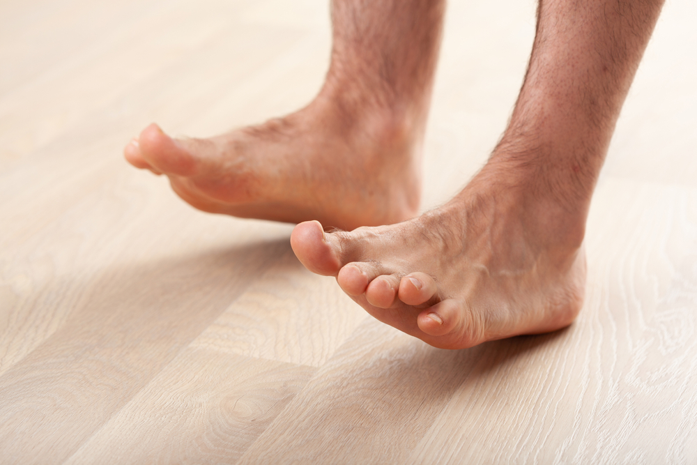
A useful home test: stand facing a wall and try to rise onto the toes of the affected foot only. Inability to do this, or significant pain with the attempt, is a strong clinical indicator of significant posterior tibial tendon involvement and warrants evaluation.
The “too many toes” sign: stand normally and look straight ahead while someone views you from behind. If more toes are visible on the outside of the affected foot than the unaffected foot, the foot has rotated outward — a visible sign of arch collapse and PTTD progression.
How Is PTTD Diagnosed?
Diagnosis starts with a thorough hands-on exam. We observe your standing alignment, check the double- and single-leg heel raise, assess arch height and hindfoot position both with and without weight bearing, evaluate the range of motion of the ankle and hindfoot, and examine the posterior tibial tendon along its course. We stage the condition based on these findings, which directly determines the treatment approach.
Imaging provides important additional detail:
Weight-bearing X-rays — standing views of the foot and ankle are essential. They show the degree of arch collapse, the alignment of the hindfoot, forefoot abduction, and any arthritis in the foot and ankle joints. Standing ankle X-rays are critical in advanced cases to identify any talar tilt that would indicate Stage IV disease.
Diagnostic ultrasound — maybeperformed in-office, ultrasound lets us see the posterior tibial tendon directly — whether it is inflamed, thickened, partially torn, or intact.
MRI — provides the most complete picture of tendon integrity, degree of tearing, and associated ligament and joint involvement. Typically ordered before surgical planning or in complex presentations.
Treatment Options at Family Foot & Ankle Clinic
Treatment is staged to match the condition. The right treatment at Stage I is different from the right treatment at Stage III. Our goal is always to arrest progression, reduce pain, and preserve function at whatever stage we first see the patient.
First-Line Conservative Care
Orthotics and bracing — the cornerstone of conservative PTTD treatment. Unlike most other conditions where orthotics are a supplement, for PTTD they are first-line treatment. The posterior tibial tendon cannot recover if it continues to be loaded without arch support. We prescribe the appropriate device based on stage:
Stage I: Custom orthotic with medial arch support and rearfoot.
Stage II: Custom orthotic plus an ankle stirrup brace for activity, or a UCBL insert for more significant arch collapse.
Stage III–IV: Arizona AFO or similar rigid ankle-foot orthosis, which controls hindfoot and ankle position when in-shoe orthotics are no longer sufficient. For Stage IV, the AFO provides the ankle stability the failed deltoid ligament can no longer supply.
Structured home exercise program. Stretching and progressive strengthening of the posterior tibial tendon — in the right sequence — are essential. We offer a complete home care program covering stretching, isometric activation, resisted inversion with a band, progressive heel raises, and doming. See our PTTD Home Care Program below.
Immobilization. For moderate Stage II or during a severe flare, a short-leg walking boot removes the load from the tendon completely, allowing the inflammation to settle before beginning rehabilitation exercises.
Activity modification. Reducing or temporarily eliminating activities that load the arch — running, prolonged standing, stair climbing — is necessary while the tendon is actively inflamed.
Footwear guidance. Motion control or stability shoes with firm heel counters and high heel-to-toe drop are the appropriate category for PTTD. Flat shoes, flip-flops, and soft-soled casual footwear worsen arch collapse with every step. See our Shoe Recommendations Guide.
Anti-inflammatory medications. NSAIDs can reduce tendon inflammation in the early stage. Used as an adjunct, not as the primary treatment.
Physical therapy. A structured PT program that includes the exercises in our home program, plus manual therapy, gait retraining, and progressive loading, accelerates recovery and is indicated for Stage II patients in particular.
Shockwave Therapy (EPAT) — Advanced Conservative Option
Shockwave therapy (EPAT). For Stage I and early Stage II PTTD where the tendon has developed chronic thickening and degeneration that is not resolving with conservative care, shockwave therapy is a meaningful option. It delivers acoustic pressure waves into the tendon to stimulate healing and break down fibrotic tissue — addressing the structural tendon changes, not just the inflammation. Because shockwave works best on chronically thickened tissue, it is more effective when used earlier in the chronic phase rather than waiting for further degeneration. It is non-invasive, no surgery, no needles, no downtime. A typical course is six sessions. Insurance does not cover shockwave; it is offered as a cash-pay service with package pricing available. → See our Shockwave Therapy (EPAT) page.
When Is Surgery Needed?
Surgery becomes necessary when conservative treatment has failed to halt progression, or when the disease has already advanced to a stage where conservative care cannot produce meaningful improvement. The earlier the stage at which surgery is performed, the simpler the procedure and the better the recovery.
See our Foot & Ankle Surgery Overview for general information on what to expect from surgical care at our clinic.
Long-Term Care & Prevention
For patients who have been treated successfully at Stage I or II, the following habits prevent recurrence and slow any further progression:
Wear your orthotics consistently — the arch mechanics that allowed PTTD to develop have not changed; the orthotic manages them
Maintain motion-control or stability footwear as the standard, not flat or unsupportive shoes
Continue calf stretching and posterior tibial tendon strengthening as a long-term maintenance program, even when pain-free
Manage weight — every pound of body weight translates to significant additional load on the posterior tibial tendon with every step
Monitor the arch and ankle alignment periodically — if the foot is changing, return for reassessment before the next stage develops
Return promptly if pain recurs — a treated Stage II patient who has a recurrence is still far easier to manage than one who progresses to Stage III
Frequently Asked Questions
What is the difference between PTTD and regular flat feet?
Flat feet can be congenital (present from childhood) or acquired (develops over time). PTTD is specifically an adult-acquired flatfoot caused by failure of the posterior tibial tendon — the tendon that was previously holding a functional arch becomes unable to do its job. This is different from someone who simply has low arches by birth. The distinction matters because PTTD is progressive and requires active treatment, while congenital flexible flatfoot is often managed with orthotics alone and does not necessarily worsen.
Can flat feet from PTTD be reversed?
In Stage I and flexible Stage II, conservative treatment can prevent further collapse and significantly reduce or eliminate pain, but it does not rebuild a lost arch. The arch support comes from the orthotic or brace, not from the tendon recovering its original structure. In Stage II surgery, procedures like tendon transfer and calcaneal osteotomy can substantially reconstruct the arch. In Stage III and IV, rigid deformity cannot be reversed — surgery corrects alignment rather than restoring a normal arch.
Can I still exercise with PTTD?
Yes, with modification. High-impact activities that repetitively load the arch — running, jumping, prolonged stair climbing — should be reduced or temporarily stopped during active inflammation. Low-impact alternatives like swimming and cycling are usually well tolerated. The strengthening exercises in our home program are specifically designed to load the tendon progressively in a way that promotes healing rather than aggravating it.
Is custom orthotics the same as a store-bought arch support?
No. A custom orthotic is molded precisely to your foot’s specific arch height, rearfoot alignment, and deformity pattern. It includes rearfoot posting that controls heel position — the key mechanical issue in PTTD. An over-the-counter arch support provides some cushioning and mild arch lift, but it cannot control the degree of rearfoot valgus that drives PTTD progression. For Stage I, a quality OTC insert may provide some relief; for Stage II and beyond, it is not sufficient.
How long does treatment take?
Stage I with consistent conservative care typically shows significant improvement within 6–8 weeks and may fully resolve over 3–6 months. Stage II is a longer process — 3–6 months of conservative management to determine whether surgical intervention is needed, and 6–12 months of recovery if surgery is performed. The single most important variable is how early treatment begins.
I’ve been told I need surgery. Should I get a second opinion?
Always reasonable — especially for Stage III and IV disease where the procedures are significant. Our Second Opinion / Foot Pain Not Improving visit is designed for exactly this situation. We will review your imaging, stage the condition independently, and give you our honest assessment of whether conservative options remain and which surgical approach makes the most sense for your specific presentation.
Arch Pain Getting Worse? Don’t Wait for It to Become a Surgical Problem.
PTTD is one of the most treatable conditions in podiatry when caught early — and one of the most difficult when caught late. Book an appointment at our Coon Rapids or Golden Valley office and we will stage your condition, tell you exactly what is going on, and build a plan that makes sense for where you are right now.
Call 763-421-7300 or Book Online
Serving Coon Rapids, Golden Valley, Maple Grove, Champlin, Blaine, Plymouth, Andover, Ham Lake, Wayzata, Minnetonka, Edina, Robbinsdale, Crystal, Brooklyn Park, and all Twin Cities communities.
Related Services & Conditions
Patient Resources
We give every PTTD patient a complete set of take-home materials at their visit. These are also available to download below: