
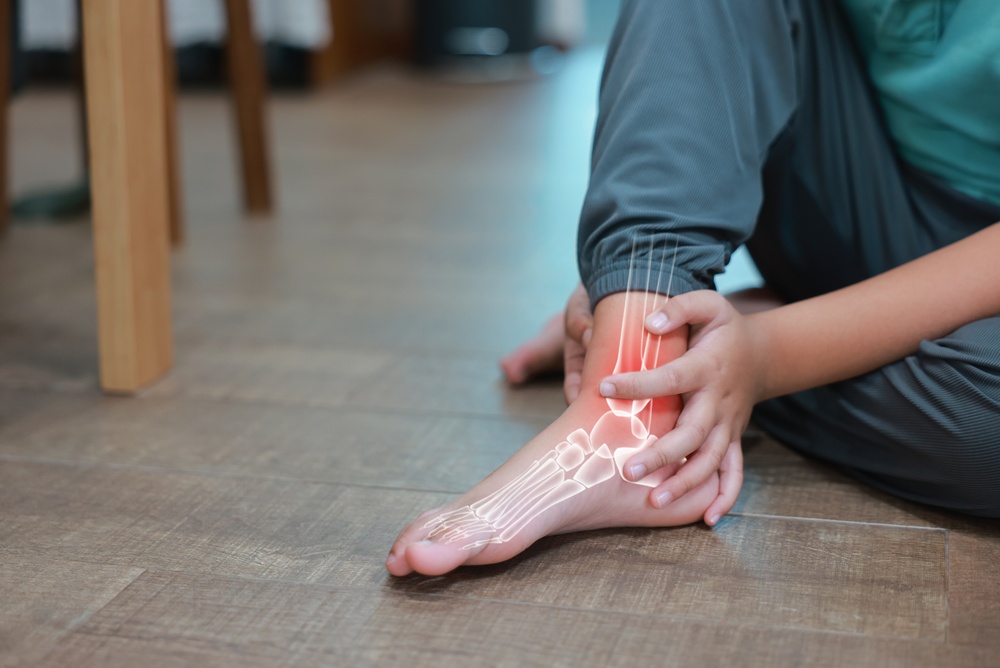
Pain That Builds With Activity and Gets Better With Rest — That Is the Pattern
A stress fracture is not the result of one traumatic event. It develops gradually as bone is subjected to repetitive loading faster than it can repair itself. The result is a spectrum of injury ranging from early bone stress reaction — where the bone is being damaged but has not yet cracked — to a complete stress fracture with a visible fracture line. What makes stress fractures frustrating is that early on they do not show on X-ray, and because the pain comes and goes with activity, many patients convince themselves it will sort itself out. It usually does not.
The foot and ankle are among the most common sites for stress fractures, particularly in runners, military recruits, dancers, basketball players, and anyone who has recently increased training load. But stress fractures are not only a sports injury — they also occur in older adults whose bone density has declined, often with minimal provocation. Our podiatrists diagnose and treat stress fractures at our Coon Rapids and Golden Valley offices, serving patients across the north and west metro including Maple Grove, Ham Lake, Andover, Champlin, Blaine, Plymouth, Wayzata, Minnetonka, St. Louis Park, and Edina.
Do not push through foot or ankle pain that worsens during activity and improves with rest. Continuing to load a stress fracture risks converting an incomplete fracture into a complete one, significantly extending recovery time and potentially requiring surgery. If the pain has been building over days to weeks rather than appearing suddenly, it needs imaging.
Call 763-421-7300 or book online
What Is a Stress Fracture?
A stress fracture is a small crack caused by repetitive loading — not a single injury. When training load increases faster than bone can adapt and repair, micro-damage accumulates until a fracture develops. Two types occur in the foot and ankle:
- Fatigue fractures: too much load on healthy bone. Typical in athletes and military recruits who increase training volume too quickly.
- Insufficiency fractures: normal load on weakened bone. Typical in older adults, postmenopausal women, and patients with osteoporosis, osteopenia, or long-term steroid use. These can occur with routine daily activity.
The distinction matters: fatigue fracture treatment focuses on load management and return to activity. Insufficiency fracture treatment also requires evaluation and management of the underlying bone health issue to prevent recurrence.
Common Sites in the Foot & Ankle
Not all stress fractures are equal. The site determines the treatment approach, the healing timeline, and the risk of complications. Stress fractures are classified as low-risk or high-risk based on the likelihood of delayed union, non-union, or progression to complete fracture with displacement.
| Site | Risk Level | Typical Patient | Key Clinical Notes |
| Metatarsal shafts (2nd, 3rd, 4th) | LOW | Runners, dancers, military recruits, patients with recent increase in walking activity | The most common foot stress fracture. Responds well to protected weight-bearing and activity modification. Rarely requires surgery. Typically heals in 6–8 weeks. |
| Fibula (lower third) | LOW | Runners, aerobics participants | Often confused with ankle sprain. Point tenderness 3–6 cm above the lateral ankle. Responds well to conservative management. |
| Calcaneus (heel bone) | LOW | Military recruits, older adults, runners | Often presents as diffuse heel pain. Must distinguish from plantar fasciitis. Squeeze test (medial-lateral compression) is positive. Good response to conservative management. |
| Distal tibia (anterior cortex) | LOW to MODERATE | Distance runners, jumpers | The anterior cortex crack can progress to complete fracture. Activity restriction and sometimes a boot required. Monitor closely. |
| Navicular | HIGH | Sprinters, basketball players, football players | Most commonly missed and most commonly complicated stress fracture in the foot. Dorsal midfoot pain. X-ray often normal — MRI or CT required. Non-weight-bearing cast for 6–8 weeks minimum. High non-union rate if undertreated. |
| Fifth metatarsal (Jones fracture zone) | HIGH | Basketball, soccer, football players; patients with cavus foot | The Jones fracture zone (proximal shaft-diaphysis junction) has poor blood supply and high non-union rate. Often requires surgical fixation with intramedullary screw, especially in athletes. Distinguish from avulsion fracture at the tuberosity (lower risk). |
| Medial malleolus | HIGH | Runners, jumpers | Relatively rare but high risk of complete fracture and non-union. Requires non-weight-bearing and often surgical fixation. Do not return to running until fully healed and cleared. |
| Sesamoids (great toe) | HIGH | Dancers, sprinters, basketball players | Stress fracture vs. bipartite sesamoid is a common diagnostic challenge. Treated conservatively with offloading in most cases, but healing is slow and incomplete fractures can progress. |
Clinical note: If you have been told you have a navicular, Jones fracture zone, medial malleolus, or sesamoid stress fracture and are not getting better, or have been given conservative treatment for more than 8 weeks without progress, this is exactly the situation for a second opinion. High-risk fractures that are undertreated can progress to complete fracture and non-union, which then require more complex surgical management than would have been needed at presentation.
What Causes Stress Fractures?
Most stress fractures result from a combination of training errors, biomechanical factors, and bone health:
- Rapid increase in training volume, intensity, or surface hardness without adequate recovery
- Worn-out or unsupportive footwear
- High-arched or flat feet, which concentrate load at specific sites
- Muscle fatigue, which transfers more impact force directly to bone
- Osteoporosis, osteopenia, or Vitamin D and calcium deficiency
- Long-term corticosteroid use, postmenopausal estrogen decline, or Female Athlete Triad / RED-S
- Prior stress fracture history — one of the strongest predictors of future fracture
Symptoms of a Stress Fracture
The hallmark pattern: pain that builds during activity and eases with rest. It starts at the end of a workout, then comes on earlier and earlier, and eventually is present with routine walking.
- Gradual onset of pain during activity that improves with rest
- Point tenderness at a very specific spot on the bone — pressing directly on the fracture site is significantly more painful than pressing nearby
- Mild swelling or warmth at the site
- Pain that is worse with weight-bearing and better at rest
Important: a normal X-ray does not rule out a stress fracture. X-rays are often negative for the first 2–3 weeks. If the clinical picture fits, an MRI is the appropriate next step.
How Are Stress Fractures Diagnosed?
Diagnosis combines the clinical history, physical exam, and imaging. We identify the fracture site, classify it as high or low risk, and determine the severity — bone stress reaction, incomplete fracture, or complete fracture — since each changes management.
- X-ray — the first step, but often normal for 2–3 weeks after a stress fracture develops. A normal X-ray does not rule out the diagnosis.
- MRI — the gold standard. Detects bone marrow edema within days of injury and shows severity. Preferred for all high-risk sites (navicular, Jones zone, sesamoid, medial malleolus).
- CT scan — best for characterizing the fracture line at high-risk sites before surgical planning.
Bone density: for any insufficiency fracture or young patient with multiple stress fractures, a DEXA scan and basic bone health labs (Vitamin D, calcium) should be ordered. Treating the fracture without addressing the underlying bone health issue results in recurrence. See our Bone Health & Fracture Prevention Guide below.
Treatment at Family Foot & Ankle Clinic
Treatment is determined primarily by the fracture site (high-risk vs. low-risk) and the degree of injury (bone stress reaction vs. incomplete vs. complete fracture). The core of treatment is protecting the bone from further loading while healing occurs.
Treatment depends on the fracture site and severity. The core principle: protect the bone from further impact loading while healing occurs.
Low-Risk Fractures — Conservative Management
- Activity modification: stop the aggravating activity. Non-impact cross-training (swimming, cycling, pool running) maintains fitness without loading the fracture site.
- Protected weight-bearing: a stiff-soled shoe, surgical shoe, or walking boot reduces bending stress at the fracture site during walking.
- Custom orthotics: corrects the biomechanical factors that caused the fracture and reduces recurrence risk. More on our Custom Orthotics page.
- Bone health and nutrition: Vitamin D and calcium are essential for fracture healing. If bone density is a concern, we coordinate with your primary care physician. See our Bone Health & Fracture Prevention Guide below for specific targets.
- Graduated return to activity: premature return is the most common cause of re-fracture. Imaging confirmation before resuming impact activity is advisable for any high-risk site.
High-Risk Fractures — More Aggressive Management
High-risk sites require non-weight-bearing and often surgical management:
- Navicular: non-weight-bearing cast 6–8 weeks minimum; CT-confirmed healing before return. Surgical fixation for athletes or failed conservative management.
- Jones fracture (proximal fifth metatarsal shaft): non-weight-bearing cast for lower-demand patients; intramedullary screw fixation for athletes due to high non-union rate.
- Medial malleolus: non-weight-bearing; surgical fixation often recommended given high non-union risk.
- Sesamoid: offloading orthotic or dancer’s pad; extended non-weight-bearing in resistant cases.
No improvement after 6–8 weeks of appropriate treatment for a high-risk fracture: this needs reassessment. Delayed non-union becomes progressively more difficult to treat — contact our office or see our Second Opinion page.
Adjunct Options for Delayed Healing
Bone growth stimulation (LIPUS): low-intensity pulsed ultrasound applied over the fracture site promotes osteoblast activity. Useful adjunct for navicular and Jones fractures managed conservatively.
Shockwave therapy (EPAT). For stress fractures taking longer than expected to heal, shockwave is an emerging option. Acoustic pressure waves stimulate blood flow and osteoblast activity at the fracture site. Non-invasive, in-office, three to six sessions. Not covered by insurance; offered as a cash-pay service. → See our Shockwave Therapy (EPAT) page.
When Is Surgery Needed?
Surgery is indicated for:
- Jones fracture zone in athletes — intramedullary screw fixation
- Navicular fracture failing 8 weeks of non-weight-bearing, or with displacement
- Medial malleolus fracture with crack propagation or displacement
- Any high-risk fracture progressing to complete displaced fracture
- Non-union at a high-risk site after adequate conservative management
See our Foot & Ankle Surgery Overview for general information on surgical care at our clinic.
Prevention
- Train gradually: increase load no more than 10% per week. Never increase volume and intensity at the same time.
- Replace footwear every 300–500 miles: shoes lose cushioning before they look worn. See our Shoe Recommendations Guide.
- Custom orthotics: essential for high-arched or flat feet that concentrate load at specific fracture-prone sites.
- Optimize bone health: Vitamin D, calcium, and a DEXA scan if you have risk factors. See our Bone Health & Fracture Prevention Guide below.
- Cross-train and rest: non-impact training between high-load days reduces cumulative bone fatigue.
Frequently Asked Questions
Can you walk on a stress fracture?
It depends on the site. Low-risk fractures (metatarsal shafts, calcaneus) often allow protected walking in a boot. High-risk sites (navicular, Jones zone, medial malleolus) typically require non-weight-bearing. Walking on an unprotected high-risk fracture can convert an incomplete fracture into a complete one. Get it evaluated first.
My X-ray was normal but I think I have a stress fracture.
This is common. X-rays are frequently negative for the first 2–3 weeks. If the clinical picture fits — activity-related pain at a specific bony point, relief with rest — an MRI is the appropriate next step. Do not wait for the X-ray to change if the suspicion is high, particularly for high-risk sites.
How long does healing take?
Low-risk sites: 6–8 weeks with appropriate protection. High-risk sites: 8–12 weeks or longer, sometimes requiring surgery. Older adults, patients with osteoporosis, and those with nutritional deficiencies typically take longer. Adherence to activity restriction is the single biggest variable.
Can I keep training while it heals?
You can maintain fitness with non-impact alternatives (swimming, cycling, pool running). Impact loading at the fracture site must stop until imaging confirms healing. Premature return is the most common cause of non-union.
My stress fracture is not healing. What are my options?
A fracture not healing on schedule — or a high-risk fracture managed conservatively without improvement — warrants reassessment. Our Second Opinion / Foot Pain Not Improving visit is designed for exactly this situation.
Foot or Ankle Pain That Gets Worse When You Exercise and Better When You Rest? Get It Evaluated.
That pattern is a stress fracture until proven otherwise. The longer a high-risk fracture goes without appropriate management, the harder it becomes to treat. Book an appointment at our Coon Rapids or Golden Valley office and we will get the right imaging, classify the injury correctly, and build the right treatment plan for where you are.
Call 763-421-7300 | Book online
Serving Coon Rapids, Golden Valley, Maple Grove, Champlin, Blaine, Plymouth, Andover, Wayzata, Minnetonka, Edina, and all Twin Cities communities.
Related Services & Conditions
- Ankle Fractures
- Broken Toe & Metatarsal Fractures
- Sesamoid Injuries
- Custom Orthotics
- Shockwave Therapy (EPAT)
- Second Opinion / Foot Pain Not Improving
Patient Resources
The following resources are available to download: